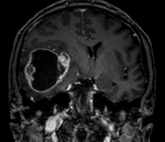
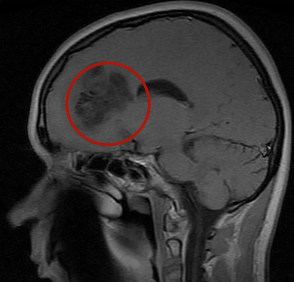
Intracerebral tumors are divided into superficial (subcortical, convexity) and deep tumors. Deep intracerebral tumors are not common, but are difficult for surgical removal due to their close location to important brain structures (thalamus, internal capsule, and other subcortical nuclei). Intracerebral tumors are localized in the frontal, temporal, parietal, or occipital lobes. Can spread to several fates at the same time. Deep intracerebral tumors include midline intracerebral tumors, intracerebral tumors that grow into the ventricular system of the brain, insular intracerebral tumors, and hippocampal/parahippocampal intracerebral tumors. According to histological distribution and degree of malignancy, tumors of the astrocytic series – astrocytoma – are distinguished (from the first to the fourth degree of malignancy, in the last case, the fourth degree, this is the most aggressive tumor – Glioblastoma – the most unfavorable prognosis). Tumors of the oligodendroglial series are distinguished: Oligodendroglioma (II-III degree of malignancy, in the last degree, the most aggressive tumor – Oligodendroblastoma). Various forms of medulloblastoma of varying degrees of aggressiveness and sensitivity to adjuvant (chemo/radiation) treatment. Separately, tumors of the pineal pocket are distinguished – pinealoma, pineoblastoma. What are the symptoms? Manifestations of intracerebral tumors are headaches, which may be accompanied by nausea/vomiting, and seizures are common. In the further course, paralysis of various degrees of severity, impaired visual acuity occurs. What is the treatment? Treatment of intracerebral tumors is combined. The surgical operation is aimed at removing the tumor, carrying out intracranial decompression, which is extremely important, and histological verification of the tumor. Chemo/radiation therapy is performed after the latter (clarification of the histology of the tumor). For surgical treatment of deeply located intracerebral tumors, tumor access is extremely important. which passes through the thickness of the brain. Minimally invasive endoscopic methods for deeply located tumors that grow into the ventricular system of the brain are the method of choice in our clinic. A clear visualization of a deeply located tumor, a narrow cerebral canal, makes it possible to activate patients more quickly, to reduce neurological deficits after surgery. In the case of deep midline intracerebral tumors, we perform microsurgical operations (using an operating microscope) if necessary with endoscopic assistance (using an endoscope to monitor possible tumor remnants. In the case of insular (islet) intracerebral tumors, we perform access through the lateral (Sylvian) In the future, we conduct lifelong monitoring (periodic MRI of the brain, periodic examinations by a neurosurgeon, if necessary, CT-oncoscreening)


