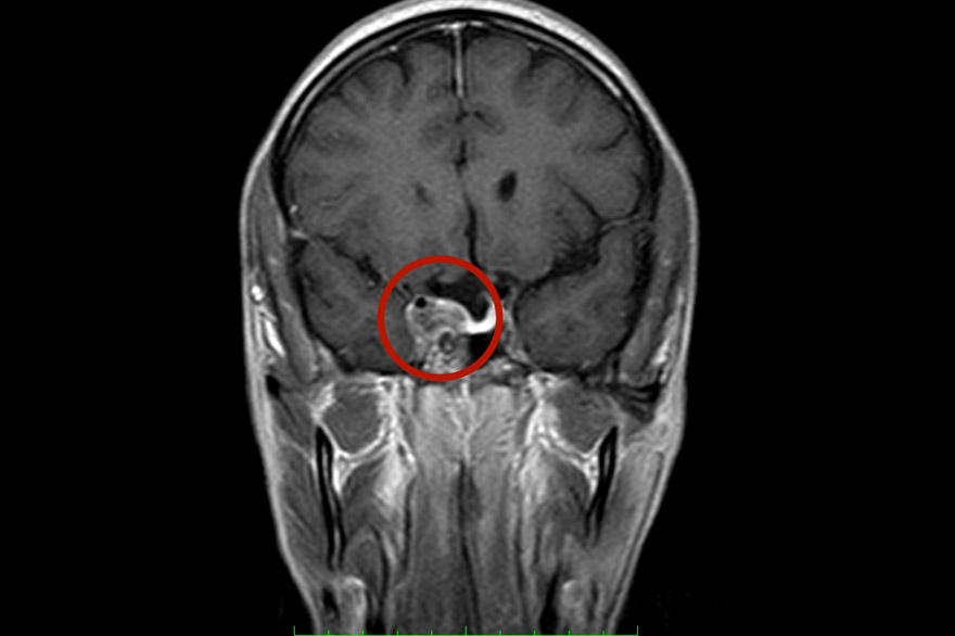
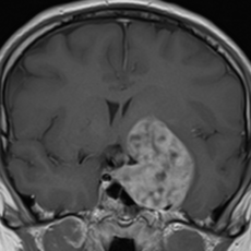
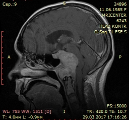
Chondroma is a benign tumor that develops from cartilage tissue. Occasionally, it can degenerate into a chondrosarcoma (less than 5%). Chondroma has slow growth, destroys the base of the skull, and only when it degenerates into a chondrosarcoma does the dura mater (which covers the brain) grow. Although chondromas acquire large sizes, the risks of surgical operation are not high (anemic tumor, with a clear plane of dissection from the dura mater).
Chondrosarcoma is characterized by a recurrent course of the disease. They are characterized by invasive growth, growing into adjacent, quite often important, structures of the base of the skull. Therefore, radical removal of chondrosarcoma is difficult. Surgical treatment can be transcranial (skull trepanation) and endoscopic endonasal. Chondrosarcomas have invasive growth, often recur and at the same time acquire intradural or subdural growth, sprouting into the dura mater of the brain.
Chordomas are tumors formed from the remnants of the notochord (the ectodermal prototype of the internal skeleton, which in the embryonic period of human development forms an axial structure) and can occur both in the base of the skull and the sacrum. The typical localization of chordoma is the area of the slope of the main bone and the cranio-vertebral junction. Chordomas have aggressive, invasive growth and over time grow into the dura mater, spreading into the posterior cranial fossa, compressing the brain stem. Chordomas have a recurrent course, after a surgical operation. Surgical interventions can be both transcranial and endoscopic endonasal, endoscopic or microsurgical transoral. The most effective radiation therapy is proton therapy.


